Knorpelschaden
Inhalt Knorpelschaden
Knorpelschaden
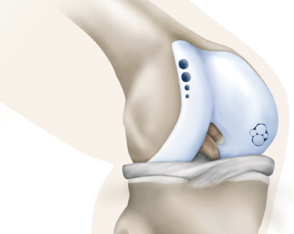
Knorpelschäden können an jedem Gelenk des menschlichen Körpers auftreten. Am häufigsten betroffen ist jedoch das Kniegelenk. Hier können Knorpelschäden zu anhaltenden Schmerzen und Schwellungszuständen führen. Insbesondere dann, wenn noch nicht das gesamte Gelenk betroffen ist, kann eine Operation zu einer anhaltenden Verbesserung der Beschwerden führen und die Ausweitung von Knorpelschäden verhindern.
Abhängig von Ort und Größe des Knorpelschadens kommen prinzipiell folgende Operationsverfahren in Frage:
Knorpeltransplantation
(Matrixgestützte autologe Chondrozytentransplantation, MACT)
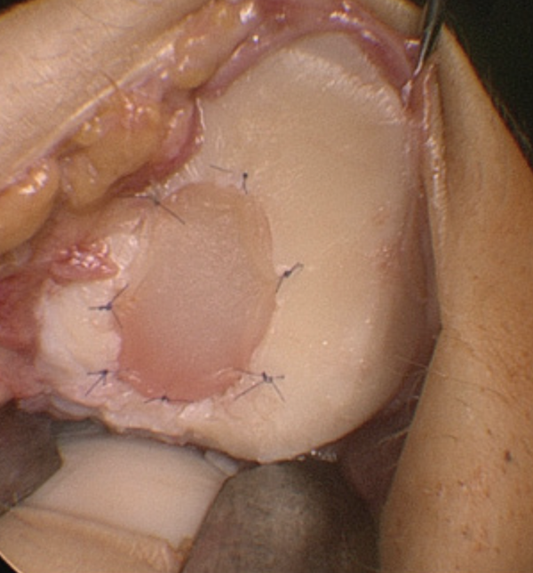
Das biologisch hochwertigste Verfahren. In einem ersten Eingriff werden körpereigene Knorpelzellen entnommen und im Labor in einer Membran angezüchtet. Diese wird bei einem zweiten Eingriff in den Knorpeldefekt eingebracht (siehe Abb. 1). Kooperationspartner des RKU ist für dieses Verfahren die Firma Tetec in Tuttlingen. Dieses Verfahren kommt für Knorpelschäden ab einer Größe von 2,5 cm² in Betracht.
Membrangestützte Knorpelgeneration
(autologe matrixinduzierte Chondrogenese, AMIC)
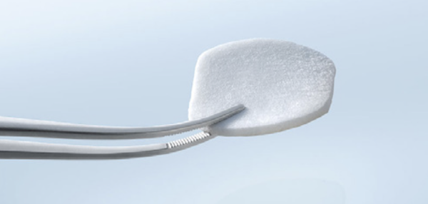
Knorpeltherapie mit Membran in einer Operation. Die Membran wird in den Knorpeldefekt eingebracht, nachdem dieser angebohrt wurde, um das Einwandern von Stammzellen zu ermöglichen (siehe Abb. 2). Die Stammzellen können langfristig neues Knorpelgewebe aufbauen.
Microfrakturierung und Pridiebohrung
(Knochenmarkstimulation)
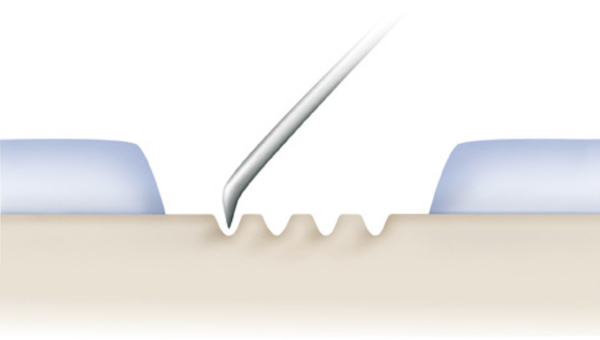
In einem Eingriff arthroskopisch anzuwenden. Der Knorpeldefekt wird präpariert und der Grund mehrfach mit einem Micromeißel (Microfrakturierung) oder einem feinen Bohrer (Pridiebohrung) perforiert, um das Einwandern von Stammzellen zu ermöglichen (siehe Abb. 3).
Autologe osteochondrale Transplantation
(OATS)
Bei kleinen Knorpelschäden. Insbesondere bei kombinierten Knorpel- und Knochenschäden werden gesunde Knorpel-Knochen-Zylinder aus einem mechanisch weniger beanspruchten Bereich in den geschädigten Bezirk versetzt (siehe Abb. 4). Der „Hebedefekt“ wird entweder mit künstlichen Knorpel-Knochen-Zylindern oder mit Knochenmaterial aufgefüllt.
Nachbehandlung
Bei jeder Art von Knorpeloperation darf der Bereich für die ersten sechs Wochen nach der Operation nicht voll belastet werden, da der „neue“ Knorpel sich erst stabilisieren bzw. bilden muss. Bis zur vollen beruflichen und/oder sportlichen Belastbarkeit dauert es bis zu einem Jahr. Detaillierte Angaben zur Nachbehandlung erhalten Sie in einem Nachbehandlungsschema. Häufig sind Kombinationseingriffe erforderlich, um z. B. den Meniskus zu reparieren oder zugrundeliegende Achsfehlstellungen oder Instabilitäten zu beheben.
Ausgewählte Publikationen
- Dornacher D, Leitz F, Kappe T, Reichel H, Faschingbauer M. The degree of correction in open-wedge high tibial osteotomy compromises bone healing: A consecutive review of 101 cases. Knee 2021; 29:478-485
- Seitz AM, Osthaus F, Schwer J, Warnecke D, Faschingbauer M, Sgroi M, Ignatius A, Dürselen L. Osteoarthritis-Related Degeneration Alters the Biomechanical Properties of Human Menisci Before the Articular Cartilage. Front Bioeng Biotechnol. 2021 May 6;9:659989.
- Sgroi M, Gninka J, Fuchs M, Seitz AM, Reichel H, Kappe T. Chondral lesions at the medial femoral condyle, meniscal degeneration, anterior cruciate ligament insufficiency, and lateral meniscal tears impair the middle-term results after arthroscopic partial meniscectomy. Knee Surg Sports Traumatol Arthrosc 2020; 28(11):3488-3496.
- Warnecke D, Balko J, Haas J, Bieger R, Leucht F, Wolf N, Schild NB, Stein SEC, Seitz AM, Ignatius A, Reichel H, Mizaikoff B, Dürselen L. Degeneration alters the biomechanical properties and structural composition of lateral human menisci. Osteoarthritis Cartilage 2020; 28(11):1482-1491.
- Sgroi M, Däxle M, Kocak S, Reichel H, Kappe T. Translation, validation, and cross-cultural adaption of the Western Ontario Meniscal Evaluation Tool (WOMET) into German. Knee Surg Sports Traumatol Arthrosc. 2018;26(8): 2332-2337
- Sgroi M, Kocak S, Reichel H, Kappe T. Comparison of 3 knee-specific quality-of-life instruments for patients with meniscal tears. Orthop J Sports Med 6 (2018) 2325967 117750082
- Dornacher D, Trubrich A, Guelke J, Reichel H, Kappe T. Evaluation of a modified knee rotation angle in MRI scans with and without trochlear dysplasia: a parameter independent of knee size and trochlear morphology. Knee Surg Sports Traumatol Arthrosc 2017;25(8): 2447-2452
- Dornacher D, Reichel H, Kappe T. Does tibial tuberosity-trochlear groove distance (TT-TG) correlate with knee size or body height? Knee Surg Sports Traumatol Arthrosc 2016; 24(9):2861-7
- Sgroi M, Faschingbauer M, Reichel H, Kappe T. Can the frontal tibiofemoral alignment be assessed on anteroposterior knee radiographs? J Orthop Traumatol 2016; 17:339-343
- Faschingbauer M, Nelitz M, Urlaub S, Reichel H, Dornacher D. Return to work and sporting activities after high tibial osteotomy. Int Orthop 2015; 39(8):1527-1534
- Joos H, Leucht F, Riegger J, Hogrefe C, Fiedler J, Dürselen L, Reichel H, Ignatius A, Brenner RE. Differential interactive effects of cartilage traumatization and blood exposure in vitro and in vivo. Am J Sports Med 2015; 43(11):2822-2832
- Nelitz M, Lippacher S, Reichel H, Dornacher D. Correlation between the classification system of Dejour and objective parameters of trochlear dysplasia. Knee Surg Sports Traumatol Arthrosc 2014; 22(1):120-127
- Lippacher S, Dreyhaupt J, Williams SR, Reichel H, Nelitz M. Reconstruction of the medial patellofemoral ligament: Clinical outcomes and return to sports. Am J Sports Med 2014; 42(7):1661-1668
- Dornacher D, Reichel H, Lippacher S. Measurement of tibial tuberosity-trochlear groove distance: Evaluation of inter- and intraobserver correlation dependent on the severity of trochlear dysplasia. Knee Surg Sports Traumatol Arthrosc 2014; 22(10):2382-2387
- Nelitz M, Williams RS, Lippacher S, Reichel H, Dornacher D. Analysis of failure and clinical outcome after successful medial patellofemoral ligament reconstruction in young patients. Int Orthop 2014; 38(11):2265-2272
- Lippacher S, Reichel H, Nelitz M. Permanent patellar dislocation after a nonoperative treatment of recurrent dislocation of the patella because of trochlear dysplasia: A case report and review of the literature. J Pediatr Orthop B 2013; 22(4):325-328
- Nelitz M, Seitz AM, Bauer J, Reichel H, Ignatius A, Dürselen L. Increasing tibial slope does not raise anterior cruciate ligament strain but decreases tibial rotation ability.. Clin Biomech (Bristol, Avon) 2013; 28(3):285-290
- Joos H, Wildner A, Hogrefe C, Reichel H, Brenner RE. Interleukin-1 beta and tumor necrosis factor alpha inhibit migration activity of chondrogenic progenitor cells from non-fibrillated osteoarthritic cartilage. Arthritis Res Ther 2013; 15(5):R119
- Lippacher S, Dejour D, Elsharkawi M, Dornacher D, Ring C, Dreyhaupt J, Reichel H, Nelitz M. Observer agreement on the Dejour trochlear dysplasia classification: A comparison on true lateral radiographs to axial magnetic resonance images. Am J Sports Med 2012; 40:837-843
- Nelitz M, Dreyhaupt J, Reichel H, Woelfle J, Lippacher S. Anatomic reconstruction of the medial patellofemoral ligament in children and adolescents with open growth plates: Surgical technique and clinical outcome. Am J Sports Med 2012; 41(1):58-63
- Nelitz M, Theile M, Dornacher D, Wölfle J, Reichel H, Lippacher S. Analysis of failed surgery for patellar instability in children with open growth plates. Knee Surg Sports Traumatol Arthrosc 2012; 20:822-828
- Lippacher S, Reichel H, Nelitz M. Radiological criteria for trochlear dysplasia in children and adolescents. J Pediatr Orthop B 2011; 20(5):341-344
- Nelitz M, Dornacher D, Dreyhaupt J, Reichel H, Lippacher S. The relation of the distal femoral physis and the medial patellofemoral ligament. Knee Surg Sports Traumtol Arthrosc 2011; 19(12):2067-2071
